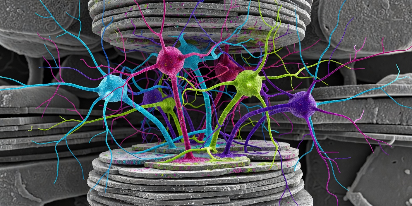
How a connectome is made
The method has not changed in principle since the 1970s. Tissue is fixed, stained with heavy metals so membranes show up, embedded in resin and cut into slices tens of nanometres thick. Each slice is imaged with an electron microscope. Then every neuron's outline is traced from slice to slice and every point where one cell contacts another with the tell-tale signs of a synapse is marked. What has changed is scale: automated microscopes, machine-learning segmentation and large teams of human proofreaders have taken the approach from a worm to a fly and now to fragments of mammalian cortex. The output is a graph — nodes and edges — plus the three-dimensional shape of every cell.
The worm, and what it taught
The first connectome was the nervous system of the nematode Caenorhabditis elegans hermaphrodite: 302 neurons and several thousand chemical synapses, published by John White, Sydney Brenner and colleagues in 1986 in the Philosophical Transactions of the Royal Society after more than a decade of hand tracing. It was extended to both sexes by Cook and colleagues in Nature in 2019. The lesson from the worm is sobering. Even with a complete map of a 302-neuron animal, predicting its behaviour from the wiring took decades of further work and still is not finished. The map generates hypotheses; it does not answer questions by itself.
The fly
The fruit fly Drosophila melanogaster was the obvious next step: a brain small enough to image entirely, complex enough to learn, navigate and court. The Janelia Research Campus released the "hemibrain" — roughly half of the central brain, about 25,000 neurons — in 2020. The larval nervous system, around 3,000 neurons, followed in Science in 2023 from Winding and colleagues at the University of Cambridge and Johns Hopkins. Then in 2024 the FlyWire consortium, led from Princeton (Sebastian Seung's group) and Cambridge and helped by hundreds of volunteer proofreaders, published in Nature the complete adult female brain: around 140,000 neurons and roughly 50 million synapses, with cell-type annotations by Philipp Schlegel and colleagues in a companion paper.
Whether a wiring diagram can predict function got its first real test the same year. Shiu and colleagues (Nature, 2024) built a simple simulation on the connectome — each neuron a leaky integrate-and-fire unit, connection weights proportional to synapse counts — and found that activating sugar-sensing neurons in the model drove the circuits that control feeding in real flies, and that predicted grooming circuits checked out. That is a modest but genuine demonstration that structure plus minimal assumptions carries some predictive power.
Mouse and human fragments
The MICrONS programme — the Allen Institute, Baylor College of Medicine and Princeton, funded through the US intelligence research agency IARPA — published in 2025, as a package of papers in Nature, the reconstruction of a cubic millimetre of mouse visual cortex: on the order of 200,000 cells and half a billion synapses. Its distinctive feature is that the same neurons were recorded with two-photon calcium imaging while the mouse watched movies before the tissue was sectioned, so anatomy and function can be matched cell by cell. That combination is what most people wanted from a connectome all along.
For humans there is H01, published in Science in 2024 by Alexander Shapson-Coe, Jeff Lichtman and colleagues at Harvard with Google Research: a cubic millimetre of temporal cortex removed during epilepsy surgery, imaged into around 1.4 petabytes of data, containing tens of thousands of cells and roughly 150 million synapses. Among the findings were rare axons forming many synapses onto a single target cell, something seldom seen in mouse. It is one sample from one person with epilepsy, so any generalisation is provisional.
The scale gap is easy to state and hard to feel. A whole human brain contains on the order of 86 billion neurons in a volume of roughly a million cubic millimetres. Imaging one cubic millimetre produced over a petabyte. A whole-brain human connectome is not a near-term project by any method now in use.
What the map structurally cannot hold
Several limits are built into the method rather than into any team's skill. Electron microscopy is a snapshot: the tissue is dead, so nothing about timing, plasticity or the state the animal was in survives. Synapse "strength" is inferred from size and count, a reasonable but imperfect proxy. Electrical synapses (gap junctions) are hard to see and often under-counted. Neuromodulators and peptides — dopamine, serotonin, the dozens of signalling molecules that act at a distance rather than across a synapse — leave no wiring trace, and in the worm their effects are known to be large. Individual variation is real: even fly brains differ between animals, so one connectome is one individual. Automated segmentation makes errors that need human correction, and the fraction corrected is a quantity every paper should state.
Where the value lies
What connectomes do well is constrain. A theory of visual cortex that requires connections the map says do not exist is in trouble. Cell-type catalogues built from morphology and connectivity line up with those from RNA sequencing, such as the BRAIN Initiative Cell Census Network atlases, and disagreements between the two are informative. Circuit hypotheses can be read straight off the graph and tested in living animals, which is what the fly community is doing at scale. That is a different kind of progress from "understanding the brain", and it is the kind that actually happens.