What the 2012 paper claimed
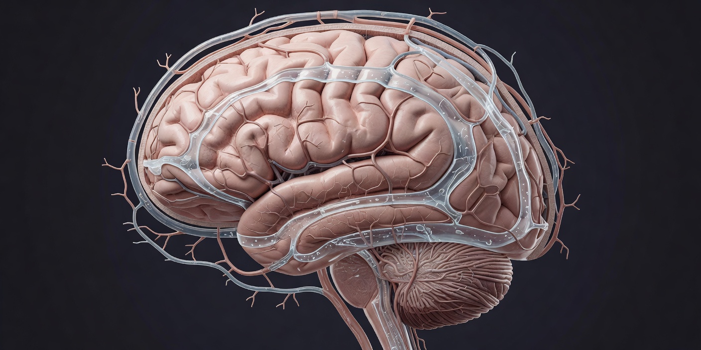
The word "glymphatic" comes from a 2012 paper in Science Translational Medicine by Jeffrey Iliff, Maiken Nedergaard and colleagues at the University of Rochester. Using two-photon microscopy in living mice, they injected fluorescent tracers into the cerebrospinal fluid and watched the tracer move into the brain along the spaces that surround arteries, pass through the tissue, and leave along the spaces around veins. The flow depended on aquaporin-4, a water channel concentrated on the endfeet of astrocytes that wrap those vessels; mice lacking the channel cleared tracer more slowly. The name joins "glia" to "lymphatic" because the brain's tissue has no conventional lymphatic vessels, and this looked like a substitute.
That much is a description of anatomy and fluid movement under specific conditions. It does not, on its own, say anything about sleep or about disease.
The sleep result
The claim that made the system famous came a year later. In Science in 2013, Lulu Xie, Nedergaard and colleagues reported that the space between cells in the mouse cortex expanded by about 60 per cent during sleep or anaesthesia compared with wakefulness, and that radiolabelled amyloid-beta — the peptide that forms plaques in Alzheimer's disease — was cleared roughly twice as fast in sleeping animals. The reasoning was tidy: a bigger interstitial space means less resistance, so fluid moves more freely and waste leaves faster. That sentence, or a version of it, is what most people now mean when they say sleep "cleans the brain".
It is worth pausing on what was measured. The clearance experiments used tracers injected directly into brain tissue, in animals that were either anaesthetised or asleep for short natural bouts, with the readout being how much tracer remained or appeared elsewhere after a set time. Each of those choices introduces a variable that a different laboratory could reasonably set differently.
What human studies can and cannot show
You cannot inject dye into a healthy volunteer's brain, so human work leans on imaging. In 2019 Nina Fultz and colleagues at Boston University published, again in Science, simultaneous EEG and fast fMRI recordings during sleep. During non-REM sleep, slow electrical waves were followed by a dip in blood volume and then by a pulse of cerebrospinal fluid into the fourth ventricle. That is a clear demonstration of coupling between brain state, blood and fluid movement. It is not a measurement of clearance; nothing was tracked leaving the brain.
Closer to a direct test is the work from Oslo University Hospital, where Geir Ringstad, Per Kristian Eide and colleagues gave patients a gadolinium contrast agent into the spinal fluid and followed it with MRI over hours and days. Contrast entered brain tissue from the surface and cleared slowly, more slowly in people with idiopathic normal pressure hydrocephalus. The limit is obvious: these are patients undergoing a clinical procedure, not a random sample, and the contrast is given by lumbar puncture rather than appearing where the brain would produce it.
Two further pieces fit the picture. In 2015 groups at the University of Virginia (Louveau and colleagues, in Nature) and the University of Helsinki (Aspelund and colleagues, in the Journal of Experimental Medicine) described lymphatic vessels in the meninges, which gives fluid leaving the brain a plausible exit. And a 2018 study in the Proceedings of the National Academy of Sciences by Shokri-Kojori, Volkow and colleagues at the US National Institutes of Health found increased amyloid signal on PET scans after a single night of sleep deprivation in healthy adults. Neither proves the glymphatic mechanism, but both are consistent with it.
The 2024 challenge
In 2024 a group at Imperial College London led by Nicholas Franks and William Wisden published in Nature Neuroscience a study that measured clearance directly in mice, injecting a fluorescent dye into brain tissue and quantifying how much remained over time. They found that clearance was lower during sleep and under anaesthesia than during wakefulness — the opposite direction from the 2013 result.
The Rochester group's response, in broad terms, was that the methods differ: direct injection of dye into tissue can itself alter the local environment, dye molecules of different sizes behave differently, and where you measure matters. The Imperial group's position is that their method is the more direct one. This is a genuine open dispute between competent laboratories, not a case of one side ignoring the other. Both agree that fluid movement changes with brain state; they disagree about whether net clearance goes up or down in sleep.
A useful way to hold this: the anatomy — perivascular spaces, aquaporin-4, meningeal lymphatics — is well supported. The coupling between sleep and fluid pulses in humans is well supported. The specific claim that sleep speeds waste removal rests mainly on one laboratory's mouse experiments and is currently contradicted by another's.
Why the mouse is a hard model here
Several trade-offs are worth spelling out. Anaesthetics are not neutral: ketamine-xylazine and isoflurane have different effects on vessel tone and on the astrocytes that matter for this system, and much of the early work used anaesthesia as a stand-in for sleep. Mice sleep in fragments across the day, so "asleep" is a short window. And a mouse brain is a few millimetres across; diffusion alone can move a small molecule that distance in a useful time, whereas a human brain would need bulk flow to achieve the same thing. Whether the human brain actually has that flow, at what rate, is not something the mouse can settle.
What would move the question on
Three things would help. A non-invasive human measure of clearance rate, rather than of fluid pulses, would let the sleep claim be tested in the species that matters. Independent replication in mice using both groups' tracers under identical anaesthesia and timing would show whether the disagreement is methodological. And longer-term studies linking measured clearance to later amyloid burden would tell us whether the mechanism has clinical weight or is a detail of fluid dynamics.
Until then, the practical position is unchanged. Sleep matters for the brain on several independent lines of evidence — memory, mood, cardiovascular risk — and none of them depends on the glymphatic model being right. Anyone with persistent sleep problems should talk to a GP rather than to a mechanism.